Fox infected with canine adenovirus-1
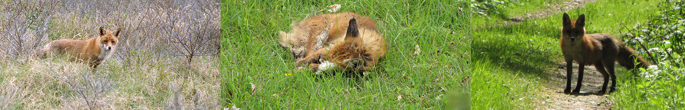
At the end of January 2024, the DWHC received a report of a dying fox by the roadside in the city Groesbeek, which was picked up by the animal ambulance of Nijmegen. The fox was taken to a veterinarian, who euthanized the suffering animal. The DWHC collected the fox for examination.
The post-mortem investigation
The fox was an adult, well-nourished female. She was not pregnant, possibly due to the presence of multiple cysts on her ovaries. The animal had no externally visible injuries, but the presence of subcutaneous haemorrhages, a ruptured liver, and blood in the abdominal cavity indicated trauma. The liver and bile duct were severely inflamed, and the fox had moderate pneumonia. Tests for Leptospirosis (Weil’s disease) and canine distemper (Canine Distemper Virus) turned out negative. The fox was then tested for adenovirus (Canine adenovirus-1, CAV-1) which was positive (PCR test).
Canine Adenovirus
The adenovirus CAV-1 is a virus found in carnivores such as canids (Canidae), bears (Ursidae), mustelids (Mustelidae), and skunks (Mephitidae).
The virus is best known as the cause of one of the classic canine diseases and is known by various names: Hepatitis Contagiosa Canis (HCC), ‘infectious hepatitis’, contagious liver disease, and Rubarth’s disease.
In unvaccinated dogs, the disease often takes a severe course, and in puppies it is often fatal. The virus can affect the liver, kidneys, and eyes of dogs, among other organs. Dogs that survive the infection can continue to shed the virus via their urine for some time, and the virus can remain infectious in the environment for a long period of time.
In the Netherlands, vaccination to protect against HCC is part of the core vaccinations that puppies receive from about 6 weeks of age, with a final vaccination at 6–12 months. The first HCC vaccination is included in the ones given around 12 weeks. After the basic vaccination, a booster is recommended every 3 years.
The CAV-1 virus has also been detected in several wild carnivore species, including fox, otter, brown bear, and skunk. Wild animals do not always show clear signs of disease, but it can sometimes be fatal for them.
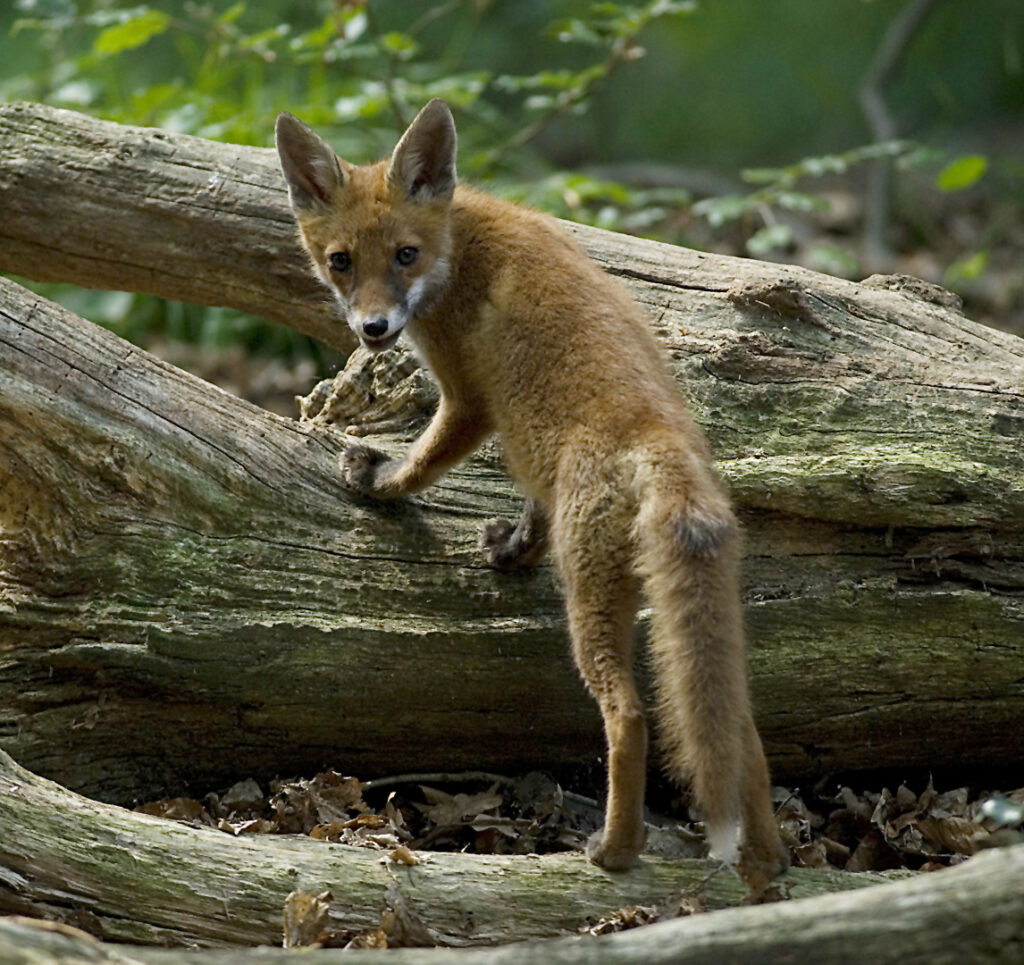
Foxes and CAV-1
It is evident that foxes can also suffer from this disease, as shown by the term found in the literature: ‘fox encephalitis’. This name was given because in foxes, mainly the central nervous system appears to be affected. However, part of the known data comes from experiments and from captive (fur) animals. The symptoms and affected organs of wild animals that have been naturally infected are relatively rarely described.
A British article from 2010 describes three cases in free-living foxes (Vulpes vulpes) in Great Britain (UK) that were brought in sick to a rescue center and died within two days.
These included a young fox in 1995 and two foxes in 2000 whose age categories are unknown. All three had (mild) jaundice; the animal from 1995 was severely weakened, and the two from 2000 were apathetic, but otherwise showed no symptoms. On necropsy, besides jaundice, a blood-rich liver and moderately enlarged liver and mesenteric lymph nodes were noted. Microscopically, extensive tissue necrosis was observed in the liver, and in the spleen and lymph nodes, a deficiency of white blood cells was noted. Typical abnormalities, so-called ‘intranuclear inclusion bodies’, were also observed in the nuclei of liver and kidney cells. The brains of these three animals were not examined. In all three foxes, a CAV-1 infection was confirmed (by virus isolation).
A more recent British article (2016) describes two outbreaks among young foxes, two to four months old, in two different rescue centres in 2011 and 2013. In 2011, four of the five fox pups housed together died within two weeks. Only some diarrhea was seen on the floor of their enclosure. In the 2013 outbreak, nine young foxes died in a short period. A few foxes showed neurological abnormalities. Four dead foxes from these outbreaks were examined. On necropsy, an enlarged liver with abnormal colour was noted, and one animal had jaundice. Microscopically, the same liver and kidney abnormalities described above were observed. Additionally, inflammation and dead cells were seen in and around blood vessels, including in the brain.
CAV-1 infection can therefore be fatal in foxes, but this is not always the case. This is shown, among other things, by a study that examined the livers and kidneys of 86 shot foxes from Italy (n=36), the UK (n=21), and Germany (n=29). The presence of CAV-1 viral material was demonstrated in the kidneys of 28% (10/36) of the foxes from Italy, 38% (8/21) of the foxes from the UK, and 3% (1/29) of the foxes from Germany. No viral material was found in the liver. The kidneys and livers from Italy and the UK were also examined microscopically, and no abnormalities indicative of disease due to CAV-1 infection (such as intranuclear inclusion bodies, liver cell necrosis, inflammation of blood vessels) were found. These findings indicate that not all infected foxes become ill and die, but that they may still excrete the virus in their urine for some time, just as dogs that have recovered from HCC do temporarily.
Scandinavian blood tests of foxes (red fox, arctic fox [Vulpes lagopus]) and wolves (Canis lupus) showed that CAV-1 antibodies are also widespread among the tested animals (respectively 59.6%, 37.8%, and 67.7%). Despite these high percentages, no disease outbreaks have been reported in these species, only occasional dead animals. This may be because dead animals are not found, because they have a high resistance and do not become ill, or because the type of virus circulating among these foxes, arctic foxes, and wolves is less pathogenic for these species.
Sources
Akerstedt, J., Lillehaug, A., Larsen, I. L., Eide, N. E., Arnemo, J. M., & Handeland, K. (2010). Serosurvey for canine distemper virus, canine adenovirus, Leptospira interrogans, and Toxoplasma gondii in free-ranging canids in Scandinavia and Svalbard. Journal of wildlife diseases, 46(2), 474–480. https://doi.org/10.7589/0090-3558-46.2.474
De Jonge, B., Van Brantegem, L., & Chiers, K. (2020). Infectious canine hepatitis, not only in the textbooks: a brief review and three case reports. Vlaams Diergeneeskundig Tijdschrift, 89(5), 284-291. DOI: 10.21825/vdt.v89i5.16956
Johnson, D. H. (2006). Miscellaneous small mammal behavior. In T. B. Bays, T. Lightfoot, & J. Mayer (Eds.), Exotic pet behavior (pp. 263–344). W.B. Saunders. https://doi.org/10.1016/B978-1-4160-0009-9.50014-1
Thompson, H., O’Keeffe, A. M., Lewis, J. C., Stocker, L. R., Laurenson, M. K., & Philbey, A. W. (2010). Infectious canine hepatitis in red foxes (Vulpes vulpes) in the United Kingdom. The Veterinary record, 166(4), 111–114. https://doi.org/10.1136/vr.b4763
Verin, R., Forzan, M., Schulze, C., Rocchigiani, G., Balboni, A., Poli, A., & Mazzei, M. (2019). Multicentric Molecular and Pathologic Study On Canine Adenovirus Type 1 in Red Foxes (Vulpes vulpes) in Three European Countries. Journal of wildlife diseases, 55(4), 935–939. https://doi.org/10.7589/2018-12-295
Walker, D., Abbondati, E., Cox, A. L., Mitchell, G. B., Pizzi, R., Sharp, C. P., & Philbey, A. W. (2016). Infectious canine hepatitis in red foxes (Vulpes vulpes) in wildlife rescue centres in the UK. The Veterinary record, 178(17), 421. https://doi.org/10.1136/vr.103559